Key Points
This guideline is intended for the use of physiotherapists utilising hyperinflation techniques as part of their routine respiratory treatments of adult, mechanically ventilated patients in ICU
Hyperinflation techniques aim to:
- increase inspiratory volume
- promote expansion of collapsed lung units
- through elastic recoil of the chest wall, help to mobilise secretions more proximally
Manual Hyperinflation (MHI) is the manual application of larger than tidal volumes, up to 2L, delivered to the patient’s invasive airway via a ‘Waters’ or Mapleson-C Circuit
Ventilator Hyperinflation (VHI) is the application of large than tidal volumes via manipulation of the ventilator settings. This can be delivered in either pressure-controlled (PC) or volume-controlled modes (VC).
VHI can be performed in SPN-CPAP/PS mode as well, but is generally not as effective as when performed in PC or VC modes.
Indications & contraindications for use are contained within this guideline appendices
This is a controlled document. If you are using a printed copy, check it against the guidelines site to ensure you are using the latest edition. Print copies must be destroyed after use.
Summary
Definitions
Hyperinflation techniques aim to increase inspiratory volume, promote expansion of collapsed lung units & in the expiratory phase, through elastic recoil of the chest wall, help to mobilise secretions more proximally (Vlaer, B. B. et al. 2021).
Hyperinflation can be performed via ETT or tracheostomy tube. It can be performed on patients who are fully sedated or have no spontaneous respiratory drive through to patients who are awake & have an intact respiratory drive. The treating therapist needs to take the patient’s underlying respiratory pattern/ drive into consideration in how they adjust their treatment. If using for secretion clearance then ideally manual techniques should be used in conjunction, unless otherwise contra-indicated (Perret, J. K. et al. 2011).
Manual Hyperinflation (MHI) is the manual application of larger than tidal volumes, up to 2L, delivered to the patient’s invasive airway via a ‘Waters’ or Mapleson-C Circuit (Maxwell, L. and Ellis, E. 2007). The technique can be adapted to include a manual inspiratory hold & a quick release of the bag to increase expiratory flow.
Ventilator Hyperinflation (VHI) is the application of larger than tidal volumes in conjunction with a prolonged inspiratory pause or hold via manipulation of the ventilator settings.
The use of chest wall shakes/ vibrations during MHI/ VHI helps to increase peak expiratory flow & to mobilise secretions.
Indications for use
Background
Hyperinflation techniques aim to increase inspiratory volume, promote expansion of collapsed alveoli and facilitate airway clearance.
Both MHI & VHI have been demonstrated to be safe & effective alternatives to standard/ usual care/ bronchial suctioning alone. Both techniques have been proven to increase pulmonary compliance & airway clearance, without adverse haemodynamic events. Additional benefits of VHI are that you can avoid disconnection from the ventilator, loss of PEEP & subsequent de-recruitment & infection risks ((Vlaer, B. B. et al. 2021).
Recent evidence also demonstrates that a more sensitive & individualised application of VHI is superior to MHI in terms of airway clearance (Jacob, W. et al. 2021). VHI has been demonstrated to significantly improve secretion clearance, compliance and oxygenation without the need for disconnecting from the ventilator & the associated risks of this (Lockstone, J. et al. 2023).
Hyperinflation techniques that are being used for secretion clearance aim to generate an expiratory flow bias to increase the net movement of secretions towards the airway (ETT or tracheostomy (Volpe, M. S., Guimaraes, M. S. and Morais, C. C. 2020).
There is evidence that VHI performed using volume-control (VC) is more likely to generate an expiratory flow bias than techniques using pressure-control (PC) or pressure-support (PS). There is also evidence that VHI protocols that utilise PIP targets of 35cmH2O are more likely to achieve an expiratory flow bias (Thomas, P. P. 2023).
In terms of lung recruitment, a minimum of 150% of the ‘ideal’ tidal volume (calculated at 8mls/kg/IBW) is required (Thomas, P. J. 2015).
The balance of achieving sufficient volumes & risk of overdistension versus expiratory flow bias must be assessed by the treating therapist in light of the aims of the specific treatment session & individual patient needs (Beatriz S. and Ribeiro, A. J. 2019).
Please see appendix 1 for a full comparison of VHI & MHI.
Risk Assessment of Treatment:
Prior to using the technique of MHI/ VHI a complete risk assessment will be undertaken, within the objective assessment, which will include the consideration of both the indications and the contraindications and potential risks of the treatment.
If it is felt MHI is the most appropriate treatment option & the patient is on high PEEP (>10 cmH2O or APRV) then consultant agreement should be sought & documented.
MHI should generally be a 2-person technique: this can be x2 physiotherapists or physiotherapist & suitable ICU healthcare professional. Ensure that the bedside nurse is aware you are about to perform MHI prior to treatment.
Physiological rationale behind hyperinflation techniques:
Please refer to above table to utilise the appropriate components of the hyperinflation technique you have chosen to best treat your patient’s problem.
Documentation
The treating therapist must fully document their rationale for & description of their hyperinflation treatment in the patient’s EPR in a timely manner. There are smartphrases for both VHI & MHI to facilitate this and can be used if the therapist so wishes.
Baseline ventilator settings & patient observations should be documented in full prior to the start & again at the end of any hyperinflation treatment.
Please ensure the response to the treatment is clearly documented.
MHI Technique:
If a patient is currently ventilated on PEEP ≥ 10cmH20 then ICU consultant agreement should be sought prior to disconnecting from the ventilator. If the PEEP is >10 consider clamping the ETT prior to disconnecting
N.B: You cannot clamp a tracheostomy
Even if the patient is sedated, you should always fully explain your treatment prior to & throughout the session.
Equipment required:
-
Filter unless one already within the ventilator circuit
-
Cuff Manometer
-
Intravenous (IV) extension line
-
Waters Circuit
-
Oxygen Supply – 15l/min
-
Keep closed suction system & EtCO2 monitor in situ
Please see appendix 4 for photographs of circuit set up
MHI Procedure:
-
Remember PIP limit of 35cmH2O during MHI
-
Ensure the Waters Circuit is connected to the oxygen, flow rate of 15 lpm
-
Aim to match the minimum pressure during expiration with the PEEP set on the ventilator
-
Silence the ventilator alarm, place ventilator on ‘Standby’ OR press the ‘manual disconnect’ button
-
Disconnect patient from the ventilator (ensuring ventilator circuit is kept clean by placing cap to end of circuit)
-
Connect patient to the Waters circuit, keep the closed suction system & EtCO2 monitor in situ
MHI with PIP target of 35cmH2O
-
Give resting breaths, keeping pressures during expiration to match PEEP set on ventilator
-
Adjust expiratory valve accordingly
-
When ready provide cycle of MHI breaths e.g.: 6-10 breaths
-
Provide slow sustained MHI breath by squeezing the Waters Circuit until either:
-
PIP target of 35cmH2O reached
-
Entire volume of Waters Circuit delivered
-
Haemodynamics become labile
-
Provide inspiratory hold for 3-5secs as tolerated, before releasing MHI breath
-
Simulate ‘huff’ by quickly releasing the ‘squeeze’ on the Water’s Circuit
-
Adjust expiratory valve if needed so as PIP of 35cmH2O not exceeded
-
Aim to keep minimum pressure within circuit during expiration, to match PEEP set on ventilator
-
Consider use of manual techniques if utilising for secretion clearance
-
Perform cycle of MHI e.g.: 6-10 breaths
-
Provide ‘rest breaths’ as described above
-
Repeat MHI cycles as required
-
Suction +/- MAC as indicated during treatment
-
Limit number of disconnections from ventilator so ideally complete all MHI treatment before reconnecting to ventilator.
VHI Technique:

VHI can be performed in many ventilator modes including: VC-AC, VC-SIMV, PC-BIPAP or SPN-CPAP/PS.
You can use VHI up to PIP/Pmax of 35cmH2O & can safely titrate your treatment to achieve the desired treatment effect up to these airway pressures.
To be considered effective VHI volumes of a minimum of 150% of 8mls/kg/IBW are required. For example if for Patient ‘A’ 8mls/kg/PIBW = is 426mls then a minimum VHI target volume would be 639mls. However, you can go higher than these minimum target volumes up to a PIP/Pmax of 35cmH2O until your desired treatment effect is achieved, as long as your patient is tolerating the treatment.
VHI has been shown to be more effective in VC modes (as compared with PC or PS modes). Consider switching from PC-BIPAP to VC-SIMV/ VC-AC for the VHI treatment.
If VHI is performed on SPN-CPAP/PS mode for secretion clearance, ideally this should be a 2-person technique so both manual inspiratory holds & manual techniques can be performed. Or else, if the physiotherapist feels competent to do so the mode can be switched to PC-BIPAP/ VC-SIMV or VC-AC (as opposed to SPN-CPAP/PS) for the duration of the VHI treatment. In these modes you can perform it as a one-person technique, including providing manual techniques for airway clearance so this should factor into your selection of ventilator mode for the treatment in order to fully utilise the inspiratory time & be able to perform manual techniques.
The expectation is that staff who are static within ICU should be competent & capable to make this assessment & switch mode as required during their treatment.
On-call staff, rotational staff, or staff new to the trust/ ICU in a static ICU role who have not yet completed all their induction competencies & training are expected to only work within their scope of practice and experience. During normal working hours they can seek the support of a more experienced ICU physiotherapist as required.
At a weekend/ Bank Holiday/ overnight on-call if there is no immediate access to a more senior ICU physiotherapist staff must work in a way which they feel is the safest for them & their patient. If this means treating the patient on the mode of ventilation on which they are currently on, then that is acceptable & that decision is fully supported by the physiotherapy department.
If these staff groups feel competent to make the assessment that another mode may be more effective for VHI but do not feel competent to make changes to the ventilator they can seek advice & practical support from the ICU NiC/ ACCP or ICU Doctors. If the VHI target volumes cannot be achieved due to hitting the PIP limits and the set PEEP is >5 cmH2O, then consideration can be given to reduce PEEP to 5cmH2O during the VHI cycles. If the treating therapist does not feel competent to make this assessment, they can seek advice from the ICU Doctors/ ACCP.
VHI cannot be performed in APRV mode. There are rare clinical situations where a patient is on APRV but may benefit from VHI for secretion clearance. In these instances, consultant agreement should be sought to change the ventilator mode from APRV to another mode of ventilation (VC-SIMV/ VC-AC/ PC-BIPAP) as appropriate for the duration of the VHI cycles only. However, there is again no expectation that staff who do not regularly work within ICU should do this without ICU support e.g.: on an overnight on-call etc. In these instances, usual physiotherapy such as positioning, manual techniques & suction is adequate.
Appendix 1: Comparison of VHI & MHI
Appendix 2: Contraindications/ precautions to hyperinflation
Appendix 3: Troubleshooting potential complications of hyperinflation
Appendix 4: MHI Circuit set up
-
Ensure Mapleson C circuit plugged into O2 & 15LO2 running
-
Ensure Expiratory valve is at least partially open
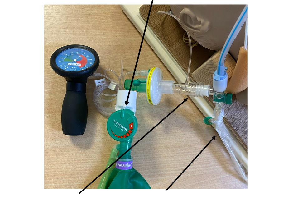
-
Keep the EtCO2 monitor and the closed suction in-line
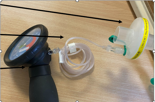
-
Ensure a bacterial filter is placed
in the circuit (if there is not one already
in the circuit), connect this to an
IV extension line, with the other end
of the extension line connected to a
manometer.
Appendix 5: How to adjust ventilator alarm settings prior to VHI on the Drager V800 ventilator - pictorial guide
- Establish minimum VHI target volume = 8mls/kg/PIBW x150%
- Find target volume of 8mls/kg/PIBW in Epic in Vent Doc Flowsheet
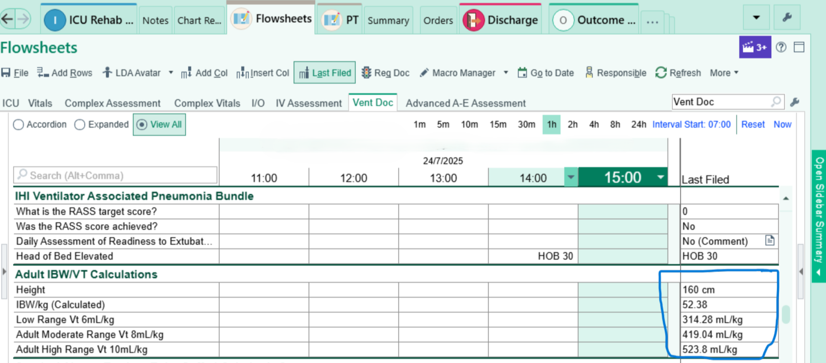
- Multiply this volume by 1.5 to get minimum VHI target VT
- Set Pmax to 35cmH2O
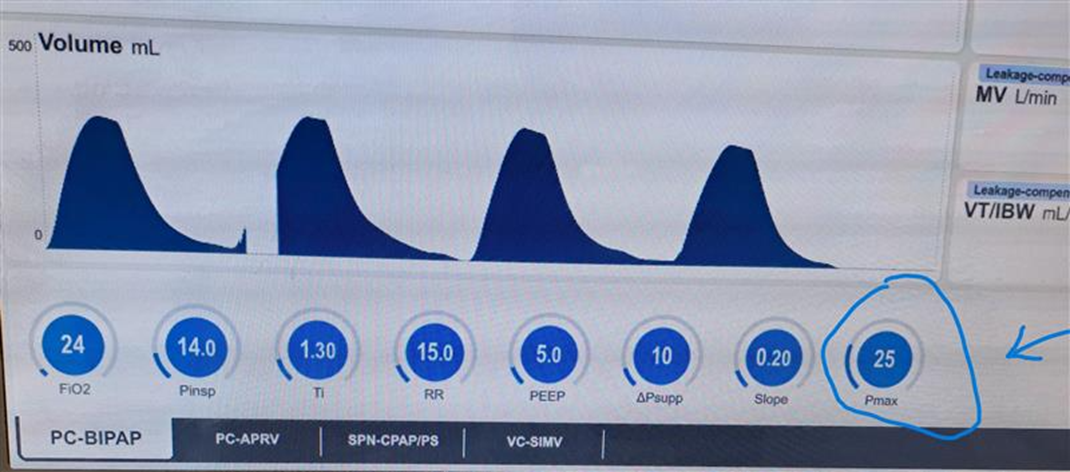
-
Set Vt upper alarm limit to 500mls above VHI target volume
-
Adjust upper MV alarm limit if needed
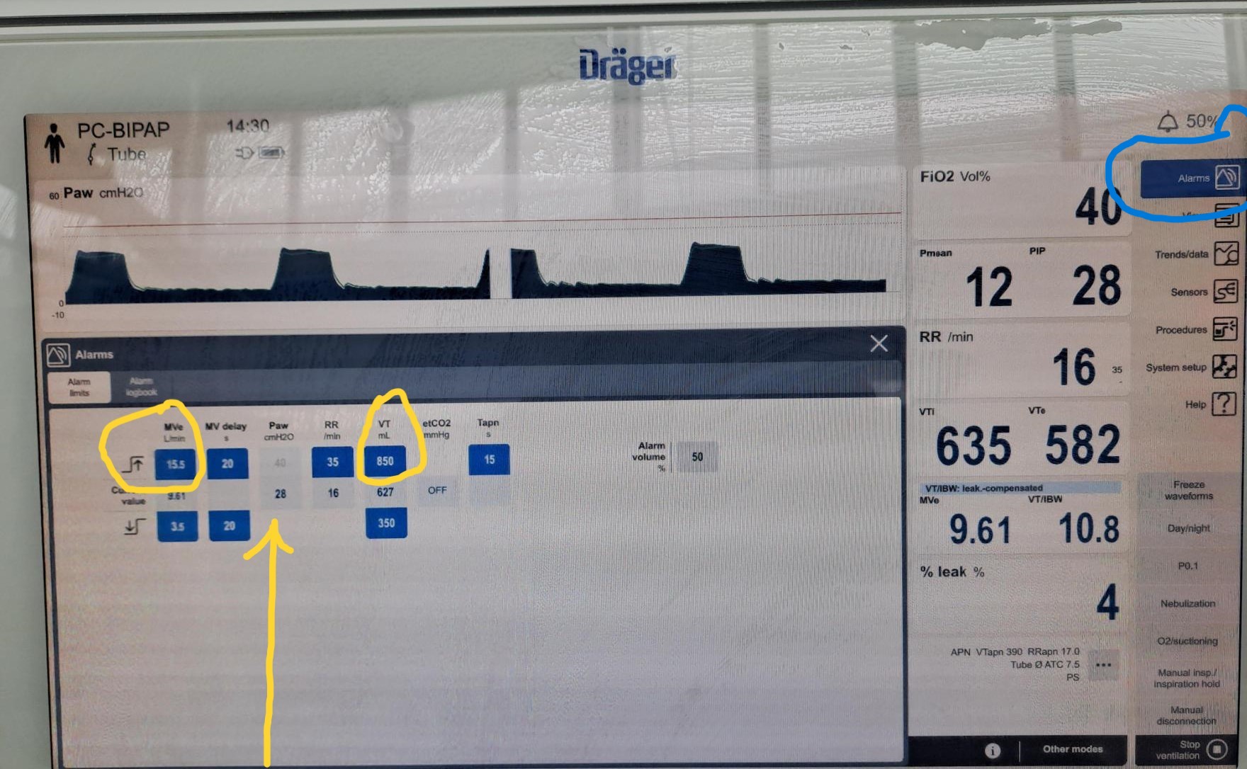
Note that the Paw upper limit is greyed out & cannot be adjusted. This is automatically set at 5cmH2O greater than the Pmax setting
Ensure you return all alarm settings to baseline once you have completed your VHI treatment session