Introduction
Hyperkalaemia is defined as an extracellular potassium ion [K+] concentration above the normal value, a serum K+ >5.5mmol/l is widely accepted.
Hyperkalaemia is a potentially life-threatening medical emergency. The incidence in hospitalised patients ranges from 1-10%. Urgent treatment can avoid life-threatening complications. Despite this, there is limited evidence to guide treatment. The Renal Association published Clinical Practice Guidelines on the Treatment of Acute Hyperkalaemia in Adults in June 2020, and more recently October 2023, which reviewed the available evidence on managing hyperkalaemia.
An important development is the availability of novel potassium binders, patiromer and sodium zirconium cyclosilicate (Lokelma), which have both been approved by NICE for the treatment of life-threatening hyperkalaemia (K+ >6.5 mmol/l), and more recently are included in the UKKA Hyperkalaemia guideline for moderate and severe hyperkalaemia. These are available for use in Frimley Health.
Factors Associated with Increased Risk of Developing Hyperkalaemia
Risk factors for Hyperkalaemia:
- Acute Kidney Injury
- Dialysis dependency (haemodialysis or peritoneal dialysis)
- Chronic Kidney Disease Stages 4 & 5 (CKD, eGFR < 30 ml/min/1.73m²)
- Drugs (renin-angiotensin-aldosterone inhibitors, NSAIDs, trimethoprim)
- Cardiac failure
- Diabetes mellitus (renin-angiotensin drugs, diabetic keto-acidosis)
- Liver disease (spironolactone, hepato-renal failure)
- Addison’s Disease (primary adrenal insufficiency)
- Hyporeninaemic hypoaldosteronism (Type IV renal tubular acidosis)
Clinical Features
Hyperkalaemia can present in numerous different ways. Symptoms are often non-specific and can often be overshadowed by other concurrent illness. Many patients are asymptomatic. In severe cases, muscle weakness or paraesthesia may occur. The clinical course is unpredictable.
Diagnosis and recognition of hyperkalaemia is normally based on laboratory tests and ECG changes.
ECG Changes
All patients presenting with hyperkalaemia should have a 12 lead ECG. When reviewing ECGs in the context of hyperkalaemia it is important to also consider co-existing metabolic disorders, e.g., metabolic acidosis and serum calcium and sodium concentrations.
Hyperkalaemia causes a rapid reduction in resting membrane potential leading to increased cardiac depolarization and muscle excitability. ECG features are present in approximately 66% of patient with severe hyperkalaemia (K+ ≥ 6.5mmol/L). The ECG may be normal, even in severe hyperkalaemia.
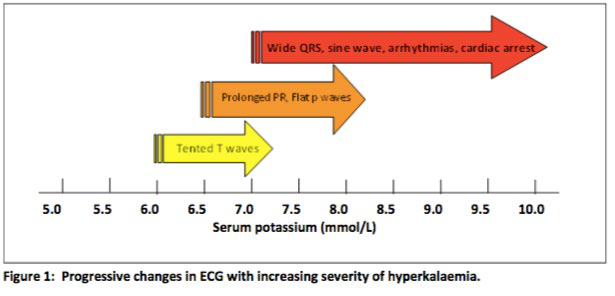
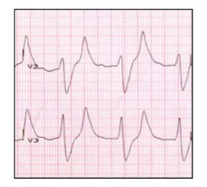
Figure 2: ECG in a patient with severe hyperkalaemia (serum K+ 9.1 mmol/l) illustrating peaked T waves (a), diminished P waves (b) and wide QRS complexes (c)
It is important to note that ECG changes do not consistently follow the step-wise pattern pictured above. ECG changes can progress rapidly. K+ concentrations >6.5 mmol/L carry a greater risk of sudden deterioration. The longer a patient has a high K+ level, the greater the risk.
Patients with pacemaker devices are not protected from the cardiac effects of hyperkalaemia. It can affect the function of both temporary and permanent pacemakers, particularly when the serum K+ exceeds 7.0 mmol/L. Hyperkalaemia causes three important clinical abnormalities in patients with pacemakers:
- Widening of the paced QRS complex
- Increased atrial and ventricular pacing thresholds that may cause failure to capture
- Increased latency manifested by a greater delay from pacemaker stimulus to onset of depolarization.
Management
Patients identified as having hyperkalaemia should be managed in an ABCDE approach. A NEWS 2 score should be completed. A thorough history identifying any risk factors for hyperkalaemia and a full drug history should be taken. The patient should be escalated to a high dependency area if there is any evidence of compromise.
If there is a reason to suspect spurious or pseudohyperkalaemia, arrange an urgent repeat venous sample for analysis on blood gas analyser.
A point of care blood gas analyser can be used to provide a rapid and reliable K+ level when an urgent result is required. A formal laboratory sample should always be sent, but treatment should be initiated if indicated based on a blood gas analyser result.
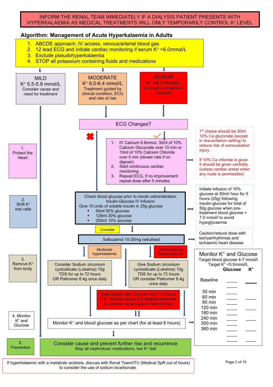
Please see Algorithm: Management of acute Hyperkalaemia in Adults (below) for treatment pathway.
Five Key Steps
There are five key steps in the treatment of hyperkalaemia. You should never walk away without completing all of these steps.
Step 1: Protect the Heart
All patients with a serum K+ ≥ 6.0mmol/L should have a 12 lead ECG. If any changes consistent with hyperkalaemia are present the patient should receive urgent treatment irrespective of the absolute K+ reading, initiate cardiac monitoring and escalate to ITU if appropriate.
Continuous 3-lead ECG monitoring should be started for all patients with a serum K+ ≥ 6.5mmol/L, features of hyperkalaemia on ECG, K+ 6.0-6.4mmol/L who are clinically unwell or who a rapid rise in K+ is anticipated. Ideally this should take place in a higher dependency setting. Cardiac stability is achieved by administering IV calcium.
Step 2: Shift K+ into Cells
Insulin is the most reliable drug for shifting K+ into cells in patients with hyperkalaemia. This effect is independent of its hypoglycaemic action. Insulin should be administered with glucose to avoid hypoglycaemia. Following insulin-glucose infusion, serum K+ starts to fall within 15 minutes, with a peak reduction occurring between 30-60 minutes. The reduction may be sustained for up to 2 hours, following which there is usually a gradual rebound. In the context of hyperglycaemia consider using an insulin only infusion.
The efficacy of insulin-glucose is increased if given in combination with nebulised salbutamol. Co-administration also appears to reduce the risk of insulin induced hypoglycaemia. The onset of action of salbutamol is within 30 minutes and duration of action is for at least 2 hours.
The main risk is of iatrogenic hypoglycaemia. Potential risk factors for this include:
- Low pre-treatment blood glucose < 7mmol/L
- Renal impairment
- Low body weight
- Older age
- Non-diabetic status
Intravenous sodium bicarbonate infusion is not used routinely for the acute treatment of hyperkalaemia.
Step 3: Remove K+ from the Body
Sodium Zirconium Cyclosilicate (Lokelma) is used as an option in the emergency management of acute life-threatening hyperkalaemia (serum K+ ≥ 6.5 mmol/l). Sodium Zirconium Cyclosilicate (SZC) is a non-absorbed potassium binder that preferentially exchanges H+ and Na+ for K+ and ammonium ions throughout the entire gastrointestinal tract. It has a rapid onset of action within 1 hour. SZC lowers K by 1.1mmol/L within 48 hours. 66% of patients will achieve normokalaemia within 24 hours.
Patiromer is a novel potassium binder. A role for patiromer in the acute setting has not been investigated as all major clinical trials were performed in stable out-patients. The onset of action is slow (4-7 hours), therefore its contribution to rapid control of serum potassium may be limited in the acute setting. Patiromer also has the potential to bind to some co-administered drugs, therefore it cannot be taken within 3 hours of other medications. However, patiromer is generally better tolerated than calcium resonium. NICE has approved patiromer for treating patients with life-threatening hyperkalaemia alongside standard medical therapy. Patiromer should not be used instead of standard treatment but could replace calcium resonium.
Calcium resonium should not be used in the emergency management of severe hyperkalaemia but may be considered in patients with moderate hyperkalaemia. Cation-exchange resins are cross-linked polymers with negatively charged structural units which entraps K+ in the distal colon in exchange for Ca2+. The most common resin used in hospitals in the UK is calcium polystyrene sulfonate (CPS), Calcium Resonium®. The onset of action is slow (> 4 hours) and efficacy is unpredictable, excluding its use in emergencies. It is also poorly tolerated due to taste and constipation. The most serious adverse effect of resins is intestinal necrosis (It is therefore no longer supported as the first line drug in acute setting). Calcium Resonium® may be considered in circumstances where patients are intolerant of other oral potassium binder and potentially in the chronic setting.
Step 4: Monitor K+ and Glucose
Serum K+ should be assessed at least 1, 2, 4, 6 and 24 hours after identification and treatment of moderate or severe hyperkalaemia, to assess efficacy of treatment and to monitor for rebound hyperkalaemia.
The peak efficacy of three of the K+-lowering drugs can be assessed at 1-2 hours. Therefore, measure serum K+ at 1 and 2 hours after initial treatment to determine if the K+ level has decreased sufficiently. Further monitoring at 4 and 6 hours is required to assess for any rebound in serum K+ as the effects of insulin-glucose and salbutamol wears off. The use of potassium binders may lower this rebound phenomenon and may provide better control of hyperkalaemia.
Measure the serum K+ at 24 hours to ensure that control of hyperkalaemia has been maintained.
Blood glucose concentration should be monitored at regular intervals (0, 30, 60, 90, 120, 180, 240, 300 and 360 minutes) after administration of insulin-glucose infusion in all patients with hyperkalaemia. Iatrogenic hypoglycaemia is a significant patient safety concern, therefore should be anticipated with regular blood glucose monitoring.
Step 5: Prevent Recurrence
Treat the underlying cause.
Ask about current medication, recent changes and use of over the counter drugs. Prescribed medication should be reviewed in the context of current illness and level of renal function. The risk of recurrence should be considered before reinstating any previous medication that may have contributed to the episode.
Drugs commonly associated with hyperkalaemia:
A low potassium diet should be started for all patients with moderate or severe hyperkalaemia.
Dialysis Patients
If a patient who has regular dialysis has hyperkalaemia escalate to the Renal team (if clinically stable and it is between 8am and 6pm) or to Intensive Care (if clinically unstable, multi organ compromise, or between 6pm and 8am).
Pseudohyperkalaemia
Results should always be interpreted with the clinical picture. Where there is discrepancy, other factors should be considered.
Situations where K+ may be falsely elevated:
- Sampling from the drip arm
- Delayed sample analysis
- Difficult venepuncture
- Sample exposed to cold temperatures
- Prolonged application of tourniquet
- Contamination with EDTA blood tube (purple top)
- Vigorous shaking
- Thrombocytosis
- Chronic Lymphocytic Leukaemia
If pseudohyperkalaemia is suspected a repeat sample in a Lithium Heparin tube (plasma) should be sent or check the K+ on the blood gas analyser. A paired clotted tube (serum) should also be sent.
Pseudo-hyperkalaemia is present if:
[Serum K+] – [Plasma K+] > 0.4mmol/L
Further Investigations
In patients presenting with hyperkalaemia U&E, a venous/arterial blood gas to assess acid base status and glucose levels are essential. In addition, it is important to consider primary adrenal insufficiency – if there is clinical suspicion, a random cortisol should be checked.
Drug charts should be reviewed in all cases of hyperkalaemia in the hospital setting. If there is concern that the hyperkalaemia is iatrogenic this must be reported on the trust incident system RL-6 for further investigation.
Drug Administration and Safety
1st choice: Calcium Gluconate – Protect the heart
Calcium Chloride – Protect the heart
Insulin and Glucose – Shift K+ into cells
Salbutamol – shift K+ into cells
Patiromer – remove K+ from the body
Sodium zirconium cyclosilicate (Lokelma) – remove K+ from the body