Key Points
-
A significant reduction or sudden alteration in fetal movements is a potentially important clinical sign.
-
Clinicians should be aware (and should advise women) that although fetal movements tend to plateau at 32 weeks of gestation, there is no reduction in the frequency of fetal movements in the late third trimester
-
From 26 weeks of gestation, all women should be asked about their fetal movements at every antenatal check.
-
Any change in pattern, reduction, or cessation of fetal movements is an indication for immediate referral to the Maternity Assessment Centre (Wexham Park Hospital) or Day Assessment Centre/ Maternity triage (Frimley Park Hospital)
-
It is important that women presenting with recurrent reduced fetal movements (RFM) are additionally informed of the association with an increased risk of stillbirth and given the option of delivery for RFM alone after 38+6 weeks.
This is a controlled document. If you are using a printed copy, check it against the guidelines site to ensure you are using the latest edition.
Print copies must be destroyed after use.
Abbreviations
Introduction
This guideline is based on the 2011 RCOG Green-top Guideline: 57 ‘Reduced Fetal Movements’, 2013 RCOG Green-top Guideline: 55 ‘Small-for-Gestational-Age: Investigations and Management, and Saving Babies Lives Care Bundle v3 2023 and Management of Reduced Fetal Movements Guideline V1, Oxford Academic Health Science Network.
(This guideline excludes the management of reduced fetal movements (RFMs) in multiple pregnancies).
The purpose of this guideline is to:
-
Ensure appropriate identification and management of acutely compromised baby
-
Prevent unnecessary iatrogenic late preterm or early term birth of a non-compromised baby
-
Aid appropriate utilisation of Antenatal ward/ Delivery Suite resources
-
Provide reassurance to staff and women that appropriate action is taken
-
Fetal movements are often perceived between 18-20 weeks of gestation and are defined as any separate kick, flutter, swish or roll. Some women may feel fetal movements as early as 16 weeks of gestation, but all should have felt movements by 24 weeks of gestation.
-
Fetal movements tend to acquire a regular pattern, and they are regarded as a significant sign of fetal wellbeing.(1)
-
A significant reduction or sudden alteration in fetal movements is a potentially important clinical sign. It has been suggested that reduced or absent fetal movements may be a warning sign of impending fetal demise.
-
Clinicians should be aware (and should advise women) that although fetal movements tend to plateau at 32 weeks of gestation, there is no reduction in the frequency of fetal movements in the late third trimester (2).
-
The number and nature of fetal movements as the fetus matures are considered to be a reflection of the normal neurological development of the fetus. From as early as 20 weeks of gestation, fetal movements show diurnal changes (3). The afternoon and evenings are often periods of peak fetal activity.
-
Fetal movements are usually absent during fetal ‘sleep’ cycles, which occur regularly throughout the day and night and usually last for 20–40 minutes. These sleep cycles rarely exceed 90 minutes in the normal, healthy fetus.
-
It has been suggested that women often feel the most fetal movements when they are lying down, fewer when sitting and fewest whilst standing.
-
Some reports suggest that 11–29% of women presenting with reduced fetal movements carry a small-for-gestational-age fetus below the 10th centile (8).
Studies have shown that 55% of women who suffer a stillbirth have perceived a reduction in fetal movements prior to diagnosis. (Several studies in the U.K and Norway have identified inappropriate responses by clinicians to maternal perception of reduced fetal movements as a common contributory factor in stillbirths) (1).
Factors which may influence women’s perception of fetal movements include
See RCOG Green Top guide 571
-
Anterior placenta - up to 28 weeks.
-
Corticosteroid administration – effects may last up to 48 hours (4)
-
Fetal position can alter perception of movements, i.e., when the fetal spine lies anteriorly to the woman.
-
Sedating drugs, such as pethidine, methadone, alcohol.
Communication and Referral
See RCOG Green Top guide 571
-
From 26 weeks of gestation, all women should be asked about their fetal movements at every antenatal check. A discussion about fetal movements and their importance should be documented. Women should be provided with the weblink for the reduced fetal movements leaflet via EPR
-
Any change in pattern, reduction, or cessation of fetal movements is an indication for immediate referral for assessment in the DAU/ MAC or maternity triage. Women should be informed to contact MAMAS line without delay if they have any concerns regarding fetal movements. They should not wait until the next day for assessment of fetal well-being.
Clinicians should be aware that instructing women to monitor fetal movements is potentially associated with increased maternal anxiety (5).
Management of RFMs before 26+0 wks (with no other symptoms present)
-
If a woman is under the care of fetal medicine, please refer to their plan of care and refer to consultant obstetrician.
-
Most women begin to feel fetal movements between 18-20 weeks of pregnancy, although multiparous women may feel them as early as 16 weeks.
-
If a woman has never felt her baby move by 24 wks, she should contact their community midwife during working hours for an antenatal check and to auscultate the fetal heart with handheld Doppler. This can be booked within one week of presentation. If required, the community midwife will make an appropriate referral to antenatal clinic.
-
Women between 16+0 and 21+6 weeks that have felt fetal movements previously and the movements are now reduced, should be advised to see their community midwife during working hours. They should be offered an antenatal check and auscultation of the fetal heart with handheld Doppler; those women should be ideally seen within 48h of presentation.
-
Women between 22+0 and 25+6 that have felt fetal movements previously and the movements are now reduced, should be advised to contact MAMAS line and will be invited to DAU/Triage/MAC for an antenatal check and auscultation of the fetal heart with handheld Doppler.
-
There is no evidence to recommend the routine use of CTG surveillance before 26+0 weeks.
-
In the event that the fetal heart cannot be auscultated in community, the woman should be referred to the Maternity Assessment Unit (MAU) for an immediate review.
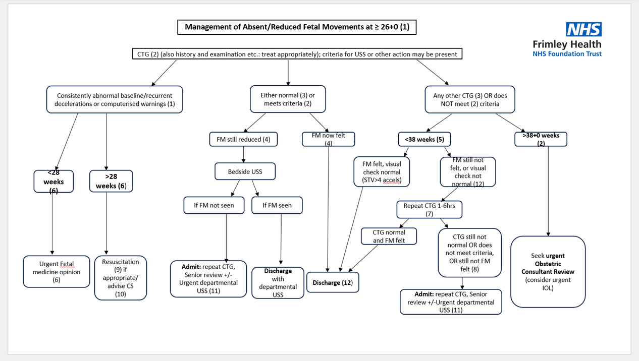
Management of RFMs after 26+0 wks (Follow flowchart 1)
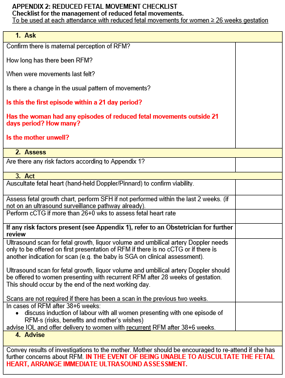
All women reporting RFMs after 26+0 wks should be invited to the DAU/MAC/ Maternity triage for further assessment. On arrival, a complete assessment of the woman should be undertaken. The Reduced Fetal Movements checklist (Appendix 2) should be filled in for each woman at each presentation with RFM.
If a woman is under the care of fetal medicine, please refer to their plan of care and refer to consultant obstetrician.
Assessment should include:
-
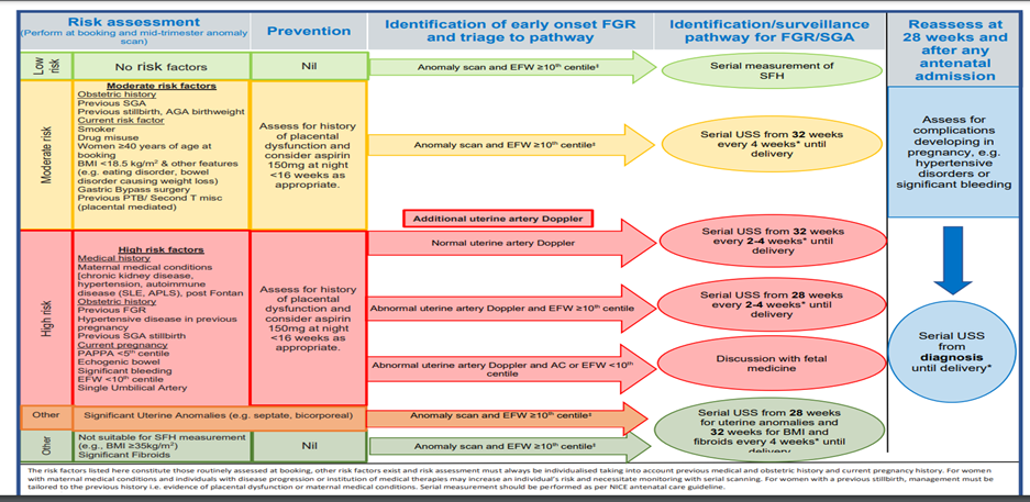
Documentation of full antenatal/medical history, including risk factors for stillbirth/fetal growth restriction according to Appendix 1.
-
Documentation of a detailed history of fetal movements.
-
Temperature, pulse and blood pressure and urinalysis
-
Abdominal palpation including symphysis fundal height measurement.
-
A cCTG (using Dawes Redman analysis) should be performed and assessed visually using the antenatal fetal monitoring analysis stickers.
-
The fetal movement button should be given to the woman so there is evidence of when she feels her baby move.
-
Discuss the importance of eating and drinking regularly.
-
Women should be reassured that 70% of pregnancies with a single episode of reduced fetal movements are uncomplicated (1).
The woman should be assessed by midwifery staff and discharged if:
-
Women are <38+6 weeks’ gestation.
-
There are no moderate or high-risk factors for stillbirth/ fetal growth restriction (see Appendix 1).
-
SFH and FH/CTG are normal
-
Women report feeling fetal movements during period of assessment.
Women should be asked to return if any further concerns regarding fetal movements. Appropriate follow-up must be in place and documented.
There is no evidence to support the use of a formal kick chart(1).
The completed checklist should be completed on EPIC and the Reduced fetal movements banner added (appears in orange colour). This is to highlight further that they have attended with a history of reduced fetal movements.
The woman should be referred for an Obstetric review and an ultrasound scan to assess for FGR should be arranged if:
-
There are moderate or high-risk factors for stillbirth or fetal growth restriction (see appendix 1) or another reason for scan (e.g., the baby is SGA on clinical assessment).
-
cCTG has not been performed.
-
The woman does not feel fetal movements during the cCTG even if the cCTG is normal.
Departmental ultrasound should be performed as soon as possible within the next working day unless they have had a scan in the previous 2 weeks. If this requires the woman to go home and return for the growth scan, then an obstetric review should be sought prior to discharge.
The healthcare professional who requests the ultrasound should indicate in the request if the woman can be discharged from the scan department in the event that the ultrasound scan is normal. If the ultrasound scan is abnormal, the woman should return to DAU/MAC/ maternity triage for obstetric review.
The woman should be referred for an Obstetric review and an Induction of labour discussed if a woman presents with a single episode of RFM after 38+6 weeks gestation.
Prior to 39 weeks’ gestation, induction of labour or Caesarean birth is associated with small increases in perinatal morbidity and neurodevelopmental delay. Thus, a recommendation for birth needs to be individualised and based upon evidence of fetal compromise (for example, abnormal CTG, EFW <10th centile or oligohydramnios) or other concerns (for example, concomitant maternal medical disease, such as hypertension or diabetes, or associated symptoms such as antepartum haemorrhage).
IOL is not recommended for a single episode of reduced fetal movements in women less than 38+6 wks and no other risk factors.
At 39 weeks’ gestation and beyond, induction of labour is not associated with an increase in caesarean birth, birth with forceps or ventouse, fetal morbidity or admission to the neonatal intensive care unit. Therefore, expediting birth by induction of labour (to women for whom this is not contraindicated) could be discussed (risks, benefits and mother’s wishes) with women presenting with a single episode of RFM after 38+6 weeks gestation.
When offering IOL, the whole clinical picture should be considered and not the history of RFMs alone. The recommendation for birth should be individualised and based upon evidence of fetal compromise (for example, abnormal CTG, EFW etc).
Management of women with continued episode of RFMs
A continued episode (re-presentation within 48 hours without normal movements between) should additionally have senior obstetric input and, if the CTG is normal, an ultrasound for fetal movements and full biometry preferably including CPR.
Management of women with recurrent episodes of RFMs
Recurrent reduced fetal movements (RFMs) can be defined as two or more episodes of RFM occurring within a 21-day period after 26 weeks, where movements have been noted in the interval. Each episode should be managed as per flowchart 1.
Additionally, SBLv3 advises ultrasound scanning (unless a scan has been normal within the last 14 days) ‘by the next working day.’ It is essential that the ultrasound findings are interpreted in the light of the clinical picture because a normal scan may occur even where a baby is severely compromised, particularly if not as a result of fetal growth restriction.
Although recurrent (but not continuous) RFMs are not strongly associated with adverse outcomes (8), it is important that women are advised of an increased risk of stillbirth and given the option of expediting birth (by the most appropriate route for them) for RFM alone after 39+0 weeks.
There is no evidence to recommend routine use of cCTG in women with normal fetal movements and normal dopplers even if reduced growth velocity is seen on growth scan. CCTG does not have predictive value in this population and should not be offered.
Auditable Standards
Women presenting with RFM after 26 weeks having a cCTG.
Women presenting with RFM after 26 weeks having a completed checklist.
Women presenting with RFM after 26 weeks with an abnormal CTG or USS.
Monitoring compliance
This guideline will be subject to three yearly audit. The audit midwife is responsible for coordinating the audit. Results presented to the department clinical audit meeting. Action plans will be monitored at the quarterly department clinical governance meeting.