Introduction
Maternal collapse is defined as an acute event involving the cardiorespiratory systems and/or central nervous system resulting in a reduced or absent conscious level (and potentially cardiac arrest and death), at any stage in pregnancy and up to 6 weeks after birth1.
The incidence of cardiac arrest in pregnancy is much rarer than maternal collapse at around 1: 36,000 maternities, with a case fatality rate of 42%. Causes of maternal cardiac arrest vary including hypovolaemia, thromboembolic disease, amniotic fluid embolism and cardiac causes with a significant mortality rate if occurring in the community. 25% of collapses were attributed to obstetric anaesthesia, all of whom were successfully resuscitated2.
An obstetric modified early warning score chart along with clinical judgment should be used to allow early recognition of the woman who is becoming critically ill1. For full guideline, see Recognising Deterioration in Pregnant or Recently Delivered Women and Use of Modified Obstetric Warning System (MEOWS).
It is essential that the clinical team are skilled in initial effective resuscitation techniques and are able to investigate and diagnose the cause of the collapse to allow appropriate, directed ongoing management 3,4.
Early involvement of senior experienced staff, including obstetricians, anaesthetists, haematologists and intensivists, is essential to optimise outcome.
2. Causes of Maternal Collapse
2.1 General
Maternal collapse can result from many causes which may or may not be pregnancy related.
A systematic approach to assessment facilitates identification of the cause of collapse. If the cause is reversible, the survival rates are greater and those for which specific treatment exists must be rapidly considered 5.
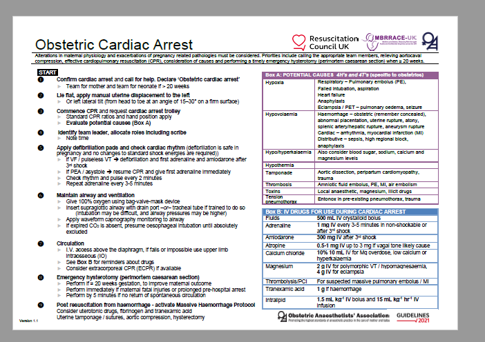
A systematic ABCDE approach should enable the clinical team to identify the most common causes of cardiac arrest. Consider 4 ‘H’s and 4 ‘T’s and, in the pregnant woman, add eclampsia and intracranial haemorrhage.
4 Hs
4 Ts
Eclampsia and pre-eclampsia
Intracranial Haemorrhage
[Resuscitation Council (UK) 2021.]
2.2 Haemorrhage
Major obstetric haemorrhage was responsible for 17 maternal deaths between 2019-2021 with an estimated incidence of 8 in 1000 maternities6. Cause of major obstetric haemorrhage include postpartum haemorrhage, major antepartum haemorrhage from placenta praevia, placental abruption, uterine rupture and ectopic pregnancy. Concealed haemorrhage should not be forgotten including following Caesarian section and ruptured ectopic pregnancy. Blood loss is often difficult to estimate and fit healthy women can tolerate significant loss prior to showing signs of decompensation1. For full guidance, refer to the Antepartum haemorrhage guideline.
In the case of maternal collapse secondary to antepartum haemorrhage, the fetus and placenta should be delivered promptly to allow control of the haemorrhage.
In the case of massive placental abruption, Caesarean section may occasionally be indicated even if the fetus is dead to allow rapid control of the haemorrhage.
Intravenous tranexamic acid significantly reduces mortality due to postpartum haemorrhage.
Point of Care ROTEM Machine should be used along with the other laboratory tests that are requested in the whole blood at the time of PPH. Its viscoelasticity technology would detect coagulation status (Platelets, Fibrinogen and Coagulation factors) and provide an overview of patient's whole blood sample in 10 minutes.
2.3 Thromboembolism
Thromboembolism is the leading cause of direct maternal death during or up to 6 weeks after the end of the pregnancy leading to the deaths of 33 women in the MBRRACE report 2019-2021.
For full guidance, refer to Thromboprophylaxis and the treatment of venous thromboembolism in pregnancy and the puerperium.
2.4 Cardiac disease
Cardiac disease remains the commonest cause of indirect maternal death and the commonest cause of maternal death overall when excluding maternal deaths from Covid-19. Between 2019 and 2021, there were 33 women that died from cardiac causes in the UK during pregnancy or within 42 days of delivery6.
The majority of deaths secondary to cardiac causes occur in women with no previous history. The main cardiac causes of maternal death are ischaemia and sudden arrhythmic cardiac death with a structurally normal heart. Aortic root dissection, although usually associated with an inherited aortopathy can present in otherwise healthy women, and signs and symptoms, such as central chest or interscapular pain, a wide pulse pressure (mainly secondary to systolic hypertension) and a new cardiac murmur, must prompt appropriate imaging and, if required, referral to a cardiologist. The incidence of congenital and rheumatic heart disease in pregnancy is increasing, secondary to increased survival rates and with improved management of congenital heart disease. In addition, women with mechanical prosthetic heart valves are at particularly increased risk of complications in pregnancy. Other cardiac causes include cardiomyopathy; dissection of the coronary artery; acute left ventricular failure; infective endocarditis; and pulmonary oedema.
After successful resuscitation, cardiac cases should be managed by an expert cardiology team.
2.5 Sepsis
Sepsis is a significant cause of maternal morbidity and mortality. Bacteraemia which can be present in the absence of pyrexia or a raised white cell count, can progress rapidly to severe sepsis and septic shock leading to collapse1.
Septic shock should be managed in accordance with the Surviving Sepsis Campaign guidelines.
For full guidance refer to the Management of sepsis in pregnancy and puerperium guideline.
2.6 Amniotic fluid Embolism
UK Incidence of Amniotic Fluid Embolism (AFE) reported in the 2019-2021 MBRRACE report was 0.39 per 100,000 maternities. AFE presents as a collapse during labour or birth, or within (usually) 30 minutes of birth in the form of hypotension, respiratory distress and acute hypoxia. Seizures and cardiac arrest may also occur1. This is followed by acute coagulopathy in those women who survive the initial event. AFE incidence is increased in women who have multiple pregnancy, polyhydramnios, placenta praevia, placental abruption and induction of labour7. If AFE is suspected or proved, the UK National Registry for AFE should be contacted.
The management of AFE is supportive rather than specific, as there is no proven effective therapy.
Point of Care ROTEM Machine should be used along with the other laboratory tests that are requested in the whole blood at the time of the event. Its viscoelasticity technology would detect coagulation status (Platelets, Fibrinogen and Coagulation factors) and provide an overview of patient's whole blood sample in 10 minutes.
Coagulopathy needs early, aggressive treatment, including the use of fresh frozen plasma.
Recombinant factor VII should only be used if coagulopathy cannot be corrected by massive blood component replacement as it has been associated with poorer outcome in women with AFE**.**
2.7 Drug toxicity and overdose
Drug toxicity and overdose should be considered in all cases of collapse. Substance misuse should be remembered as a potential cause of collapse, especially outside of hospital.
Therapeutic drug toxicity is possible with commonly used drugs in obstetric practice such as magnesium sulphate in the presence of renal impairment and local anaesthetic agents. The antidote to cardiac dysrhythmias, respiratory depression or resistant hypotension caused by magnesium toxicity is 6.8mmol of 10% calcium gluconate (30ml if weight >50kg) or 10% 6.8mmol 10% calcium chloride (10ml if weight > 50kg) given by slow intravenous injection over 5-10minutes. For full information, please refer to Toxbase®.
Signs and symptoms of Local Anaesthetic Toxicity
If local anaesthetic toxicity is suspected, stop injecting immediately.
Lipid rescue should be used in cases of collapse secondary to local anaesthetic toxicity.
Intralipid 20% is available on the Resuscitation trolley.
Manage arrhythmias as usual, recognising that they may be very refractory to treatment.
All cases of lipid rescue should be reported to NHS Improvement and the Lipid Rescue site.
Refer to the guideline for Management of severe local anaesthetic toxicity (Royal College of Anaesthetists).
2.8 Eclampsia
Fitting (Seizures) after 20 weeks gestation may be attributed to eclampsia, notably where there is no known history of epilepsy. However, epilepsy should always be considered in cases of maternal collapse associated with seizure activity.
For full guidance refer to the Hypertensive disorders in pregnancy guideline.
2.9 Intracranial haemorrhage
Intracranial haemorrhage is a potential complication of uncontrolled, particularly systolic hypertension, but can also result from ruptured aneurysms and arteriovenous malformations. The initial presentation may be maternal collapse, but often severe headache precedes this. Neuroradiologists and neurosurgeons should be involved in the care of pregnant women with intracranial haemorrhage at the earliest opportunity.
2.10 Anaphylaxis
Anaphylaxis is a severe, life threatening systemic hypersensitivity reaction, resulting in respiratory, cutaneous and circulatory changes, and collapse. There is significant intravascular volume redistribution, which can lead to decreased cardiac output. Acute ventricular failure and myocardial ischaemia may occur.
Airway obstruction secondary to angioedema, bronchospasm and mucous plugging of smaller airways all contributing to significant hypoxia and difficulties with ventilation1.
In cases of anaphylaxis, all potential causative agents should be removed, and the ABCDE approach to assessment and resuscitation followed.
If a cardiac arrest secondary to an anaphylactic reaction occurs in the community, the woman should have basic life support, including IM adrenaline if available, and be transferred to a hospital setting as quickly as possible, unless a suitably trained healthcare professional is present with appropriate equipment and drugs in which case definitive resuscitation and treatment should be commenced.
The treatment for anaphylaxis is 1:1000 (1mg/ml) adrenaline injection: Give 500 micrograms (0.5 ml) intramuscularly. This dose is for intramuscular use only.
3.0 Physiological and anatomical changes in pregnancy that affect resuscitation.
It is essential that anyone involved in the resuscitation of pregnant women is aware of the physiological differences.
Aortocaval compression significantly reduces cardiac output from 20 weeks of gestation onwards and the efficacy of chest compressions during resuscitation.
Changes in lung function, diaphragmatic splinting and increased oxygen consumption make pregnant women become hypoxic more readily and make ventilation more difficult.
Difficult intubation is more likely in pregnancy. Weight gain in pregnancy, large breasts inhibiting the working space and laryngeal oedema can all contribute to making intubation more difficult.
Pregnant women are also at an increased risk of aspiration.
4.0 Management of the collapsed woman
Maternal collapse resuscitation should follow the Resuscitation Council (UK) 2021 guidelines using the standard ABCDE approach, with some modifications for maternal physiology, in particular relief of aortocaval compression.
4.1 In the event of maternal collapse
All sample should be processed as URGENT by Pathology.
Ongoing treatment will depend on the cause of the collapse.
-
If No response
- Call 2222 for Maternal Cardiac Arrest
- Ensure manual uterine displacement in women above 20 weeks gestation or where the uterus is palpable at or above the level of the umbilicus.
4.2 Airway
-
Open airway
-
Check for obstruction
-
Head tilt, chin lift
-
Assess for breathing for up to 10 seconds
- Look for chest movement, listen for breath sounds, feel for air
-
Breathing normally
-
Turn into recovery position
-
Check help is on the way
-
Assess breathing, pulse, blood pressure, fetal heart rate regularly using a MEOWS chart.
-
Not breathing , start CardioPulmonary Resuscitation (CPR) as per Resuscitation Council (UK) 2021 guidelines
The airway should be managed as soon as possible by an experienced anaesthetist.
Bag and mask ventilation should be undertaken until intubation
- When the defibrillator arrives, apply self-adhesive defibrillation pads to the woman and analyse the rhythm. These should be applied whilst chest compressions are ongoing.
4.3 Circulation
-
During CPR
-
Establish IO/ IV access – ideally two wide bore cannulae should be inserted as soon as possible. If peripheral venous access is not possible early consideration of intraosseous access (IO), central venous access or venous cutdown should be considered.
-
Give adrenaline as per Advanced Life support algorithm.
-
Correct reversible causes – 4 H’s, 4T’s (see above, section 2.1)
4.4 Perimortem Caesarean Section (PMCS)
-
PMCS should be seen as a resuscitative procedure to be performed primarily in the interests of maternal survival.
-
Senior staff should be involved at an early stage.
-
In women over 20 weeks of gestation, if there is no response to correctly performed CPR within 4 minutes of maternal collapse or if resuscitation is continued beyond this, then PMCS should be undertaken to assist maternal resuscitation. Ideally, this should be achieved within 5 minutes of the collapse.
-
A scalpel and umbilical cord clamps should be available on the resuscitation trolley in all areas where maternal collapse may occur, including accident and emergency department.
-
Perimortem caesarean section should be performed where the resuscitation is taking place.
-
The operator should use the incision which will facilitate the most rapid access. This may be a midline vertical incision or a suprapubic transverse incision.
-
Where the outcome is not successful, the case should be discussed with the coroner to determine whether a post-mortem is required before any medical devices such as lines and endotracheal tubes are removed, as per the Royal College of Pathologists recommendations.
7.0 References
1. Chu J, Johnston TA, Geoghegan J, on behalf of the Royal College of Obstetricians and Gynaecologists. Maternal collapse in Pregnancy and the Puerperium. BJOG 2020;127:e14-e52
2. Beckett VA, Knight M, Sharpe P. The CAPS Study: Incidence, management and outcomes of cardiac arrest in pregnancy in the UK: a prospective, descriptive study. BJOG 2017; 124:1374-81
3. Healthcare Improvement Scotland. Scottish Confidential Audit of Severe Maternal Morbidity: reducing avoidable harm. 10th Annual Report Edinburgh: HIS 2014.
4. Manning E, O’Farrell IB, Corcoran P, de Foubert P, Drummond L, McKernan J, et al. Severe Maternal Morbidity in Ireland Annual Report 2015. Cork: National Perinatal Epidemiology Centre; 2017
5. Resuscitation Council UK (2021) Adult advanced life support. https://www.resus.org.uk/library/2021-resuscitation-guidelines/adult-advanced-life-support-guidelines
6. Knight M, Bunch K, Tuffnell D, Shakespeare J, Kotnis R, Kenyon S, Kurinczuk J. Saving Lives, Improving mother’s care. Lessons learned to inform Maternity Care from the UK and Ireland Confidential Enquiries into Maternal Deaths and Morbidity 2019-2021. Oxford: National Perinatal Epidemiology Unit, University of Oxford 2023.
7. Fitzpatrick K.E., Van Den Akker T, Bloemenkamp K.W.M, Risk factors, management and outcomes of Amniotic Fluid Embolism: A Multicountry population-based cohort and nested case control study. PLOS Medicine Nov 2019