Key Points
-
Endotracheal suctioning (ETS) is a common intervention performed in patients with an artificial airway.
-
There are many associated risks and complications of ETS.
-
It is an invasive procedure and should only be carried out if indicated by a competent healthcare professional.
This is a controlled document. If you are using a printed copy, check it against the guidelines site to ensure you are using the latest edition.
Print copies must be destroyed after use.
Abbreviations
1.0 Introduction
Endotracheal suctioning (ETS) is a common intervention performed in patients with an artificial airway. It is a procedure that involves the mechanical aspiration of pulmonary secretions from a patient’s airway to prevent obstruction; it is also used to maintain airway patency and prevent complications related to retention of secretions.
There are many associated risks and complications of ETS. The recommendations prior to suctioning include a comprehensive patient assessment and patient preparation. The recommendations during suctioning include appropriate catheter selection, depth of insertion, suction pressure, duration of procedure and number of suction passes. Prevention of infection and maintenance of asepsis i.e. hand washing, wearing gloves, apron and safety glasses are also essential. Suctioning is an invasive procedure and should only be carried out if indicated and not on a routine basis.
Critical Care patients that are intubated may have increased mucous production due to a respiratory infection or disease process. These patients are less able to clear secretions due to sedation or poor physiological reserve which may inhibit a normal cough reflex, increasing the risk of secretion retention and mucous plugging.
ETS can be distressing and painful for intubated patients therefore, careful selection of analgesia and adequate sedation should be considered prior to performing the procedure. ETS requirements should be assessed each shift by the health care professional. Frequency of suctioning should be guided by the viscosity of secretions, change in respiratory parameters and airway status. It is also recommended that the grading of intubation and length at patient teeth/lips is checked prior to ETS and documented appropriately in EPIC.
Any difficulty in passing a suction catheter may indicate that the Endotracheal Tube could be partially blocked and requires immediate medical attention.
2.0 Anatomy and Physiology
The anatomy of the respiratory system consists of the upper and lower respiratory tract structures.
The upper respiratory tract structures include: mouth, nasal cavities and pharynx.
The lower respiratory tract structures include: larynx, trachea, main bronchus, bronchioles and alveoli (see Figure 1).
The respiratory system has two main functions:
Inhaled air is warmed and filtered from the nose to the bronchioles and then into the alveoli and capillaries to perform gaseous exchange.
In a healthy adult inspired particles are breathed in from atmospheric air which can be cleared on coughing and expiration. Goblet cells produce mucous which line the respiratory tract and assist in removal of particles. The upper respiratory tract mucous material is moved by cells down to the pharynx. In the lower respiratory tract, the mucus moves up towards the pharynx. Mucous cells are removed by macrophages in the bronchioles, these secretions are then pushed to the oropharynx where they are expectorated or swallowed. If these individual processes are compromised, blocked or damaged it can lead to respiratory failure.
Figure 1 Anterior view showing organs of respiration
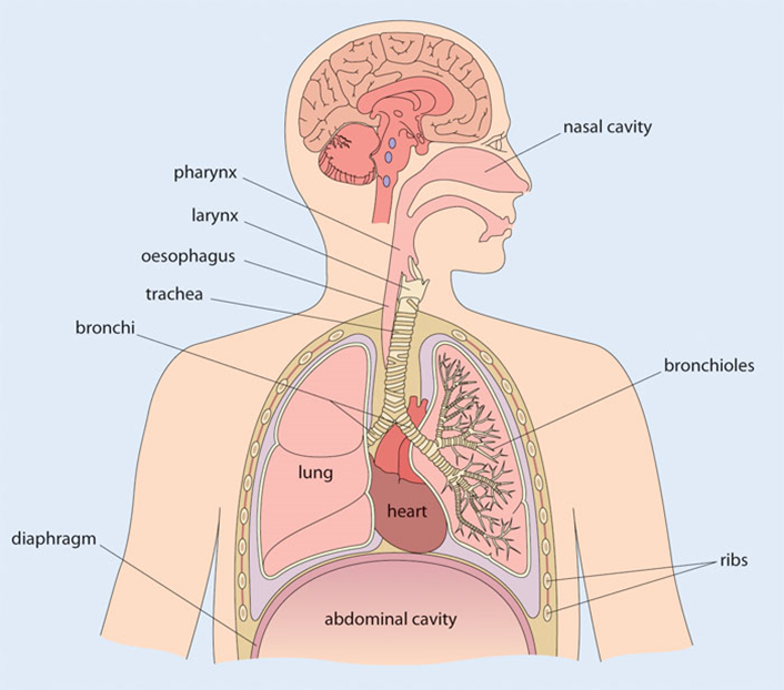
Source: Totora & Derrickson (2011)
3.0 Purpose
Suctioning of the respiratory tract involves the use of a flexible sheathed suction catheter which is connected to a suction unit that produces negative pressure. Ventilatory support continues during the procedure and is often used in patients that are receiving increased support. Evidence suggests that the suction pressure should not exceed greater than 20 KPa.
There are two current methods of ETS open and closed. The open suctioning technique involves disconnecting the patient from the ventilator, while the closed technique involves attachment of a sterile, closed in line suction catheter to the ventilator circuit. This allows the advancement of a suction catheter through the patients’ artificial airway. Disconnection from the ventilator is not required using this method. Current research has shown that this is the preferred, most common technique. The closed suction technique reduces the risk of infection to the patient and health care professional.
It prevents the loss of Positive End Expiratory Pressure (PEEP) and loss of lung volume. Research also recommends that an Endotracheal Tube (ETT) with a subglottic port reduces the risk of ventilator associated pneumonia (VAP). The subglottic port allows the nurse to aspirate secretions from around the ETT cuff preventing the risk of bacterial translocation.
4.0 Indications for suctioning
The decision to suction should be based on individual patient assessment. Suctioning should be done as rarely as possible and as frequently as needed (see section 5, Precautions and complications).
5.0 Precautions and complications to consider when performing ETS
Respiratory
-
Hypoxia/hypoxaemia
-
Bronchoconstriction/bronchospasm
-
Tracheobronchial mucosal trauma
-
Atelectasis (alveoli collapse)
-
Haemodynamic instability e.g. (bradycardia due to vagus nerve stimulation)
-
Pulmonary oedema
-
Pulmonary haemorrhage
-
Unplanned extubation
-
Right upper lobe collapse (excessive suction pressures)
-
Pneumothorax
NB: These conditions may be exacerbated by suctioning and extra precautions should be taken.
Cardiovascular
Neurological
6.0 Suctioning should be considered when:
-
There are audible coarse crackles on auscultation
-
There are audible sounds on inspiration
-
Reduced tidal volumes (if on pressure-controlled ventilation)
-
Increased airway pressure
-
A saw tooth pattern on the flow-volume on the ventilator screen indicating secretion retention
-
Reduced chest movement or breath sounds
-
Decrease in oxygen saturation (SаO2)
-
Arterial blood gases (ABG’s) indicate an increased PаCO2 or a reduction in PаO2
-
There is evidence of central and peripheral cyanosis on patient observation
-
Visible or audible secretions – rattling or bubbling sounds, audible with or without a stethoscope
-
Bradycardia / tachycardia
-
Changes in respiratory rate and pattern with increased respiratory distress
-
Suspected endotracheal tube obstruction
-
Ventilator alarms i.e. Increased airway pressure / decreased tidal volume
-
Decreased chest excursion / asymmetry
-
Patient agitation
7.0 Equipment
-
Oxygen source
-
Monitoring equipment – oxygen saturation, heart rate and blood pressure
-
Suction apparatus
-
Appropriately sized suction catheter
-
Selection of clean disposable gloves
-
Disposable plastic apron
-
Safety glasses
-
Alcohol gel
-
Sterile Water for Irrigation
Evidence suggests that suctioning a patient for no longer than 10-15 seconds is a good general recommendation. A longer duration of suction can increase negative pressure within the lungs and reduce lung volumes. It can also increase the risk of hypoxia with associated complications.
8.0 Preoxygenation prior to ETS
In general, studies have not yet shown the optimal level or duration for the use of preoxygenation for ETS. In adults, the common practice is to preoxygenate with 100% oxygen before, during and for some period after performing the procedure. Evidence suggests prolonged hyperoxia may lead to free-radical tissue damage, absorption atelectasis (nitrogen washout) and loss of lung volume. Another concern is hyperoxia in patients with cardiac related issues for example cyanotic heart disease, where hyperoxia may cause pulmonary vascular dilation and decreased preload to the left heart resulting in systemic hypotension (1).
If a patient is already hypoxic prior to ETS preoxygenation may be worth considering. Seek advice from multidisciplinary team:
ICU Doctor
Advanced Practitioners
Nurse in Charge (NIC)
Senior nursing staff
Physiotherapist
9.0 Normal Saline instillation
There is general acceptance that normal saline instillation (NSI) lubricates the ETT so that the suction catheter can pass more easily, dilutes secretions and helps to stimulate a cough. Research, however, has shown that normal saline and mucus do not mix together. There is some reservation that NSI thins or loosens secretions and improves secretion return. The common factor in the use of NSI is that it can cause desaturation and bradycardia and may increase airway infection. There have been a number of studies comparing ETS with and without NSI which show that both have the same effect upon heart rate, oxygenation and blood pressure. Another factor to consider is that NSI may cause dispersion of secretions into other areas of the lungs, or possibly lung contamination from biofilm microbes within the ETT lumen Therefore, on admission a patient should have a new in line suction catheter connected prior to performing a bronchial lavage (BAL).
Normal saline installation should not be routinely used for ETS, however adequate airway humidification is essential which is a good general recommendation.
10.0 Suction Catheter Size
If a suction catheter is too large for an endotracheal tracheal tube (ETT) and/or there is too much vacuum pressure, atelectasis may occur.
Select the correct size catheter. As a guide, the diameter of the suction catheter should not exceed one half of the internal diameter or fifty percent of the tracheal tube. This then allows air to enter the lung on suctioning, reducing the risk of atelectasis caused by increased negative pressure.
11.0 The following formula can be used to determine the correct size catheter
Suction catheter size (Fr) = 2 X (size of ETT) -2
Example: Size 8.0mm ETT X 2 (– 2) = 14f
12.0 ETS Problem solving guide
13.0 Preparation and Assessment
Note: See local competency and National Competency document for suctioning via an ETT/tracheostomy tube using closed and open suction technique.
Step 1 National Competency Page 16 1:2.3
References
-
Hahn, M. (2010) 10 Considerations for Endotracheal Suctioning. [online] Available from: http://www.rtmagazine.com/2010/07/10-considerations-for-endotracheal-suctioning/
-
American Association of Respiratory Care (AARC) (2010). Endotracheal Suctioning of Mechanically Ventilated Patients with Artificial Airways. Respiratory Care Vol 55:6 758-764.
-
Celik, S.S. and Elbas, N.O. (2000). The Standards of suction for patients undergoing endotracheal intubation. Intensive and Critical Care Nursing 16: 191-198.
-
Halm, M.A. and Krisko-Hagel, K. (2008). Instilling normal saline with suctioning: beneficial technique or potentially harmful sacred cow? American Journal of Critical Care 17: 469-472.
-
Jongerden I.P, Kesecioglu, J. et al. (2012). Changes in heart rate, mean arterial pressure, and oxygen saturation after open and closed endotracheal suctioning: A prospective observational study. Journal of Critical Care. 27 647-654.
-
Jongerden I.P, Maroeska, M., et al. (2007). Open and closed endotracheal suction systems in mechanically ventilated intensive care patients: A meta-analysis. Critical Care Medicine. 35:1 260-270
-
Leddy R, Wilkinson J.M. (2015). Endotracheal Suctioning Practices of Nurses and Respiratory Therapists: How well do they align with clinical practice guidelines? Can J Respiratory Therapy 51:3 60-64.
-
Salvatore, M.M. Lellouche, F. Pignataro., et al. (2013). Decreasing the adverse effects of endotracheal suctioning during mechanical ventilation by changing practice. Respiratory Care 58:10 1588-1596.
-
Mallett, J. Albarran, J.W, Richardson A. (2013). Critical Care Manual of Clinical Procedures and Competencies. Chichester: Wiley Blackwell pp.91-93.
-
Moore T. (2003) Suctioning techniques for the removal of respiratory secretions. Nursing Standard 18(9): 47-55.
-
Niel Wise B, Snoeren R, Van Den Broek P. (2007). Policies for endotracheal suctioning of patients receiving mechanical ventilation: a systematic review of randomised controlled trials. Infection Control Hospital Epidemiol. 28: 531-536.
-
Nursing and Midwifery Council (NMC) (2015). The Code: Professional Standards of Practice and Behaviour for Nurses and Midwives. NMC pp.9.
-
Pedersen C, Rosendahl-Nielsen, Hjermind J, Egerod I. (2009). Endotracheal suctioning of the adult intubated patient – what is the evidence? Intensive Critical Care Nursing; 25:21-30.
-
Russian, C.J, Gonzales J.F, Henry, N.R (2014). Suction catheter size: An Assessment and Comparison of 3 Different Calculation Methods. Journal of Respiratory Care 59:132-38.
-
Totora, G.J. and Derrickson, B.H. (2023) Principles of Anatomy and Physiology. 16th international adaptation edn. Hoboken: John Wiley and Sons.
Full version control record
This guideline has been registered with the Trust. However, clinical guidelines are guidelines only. The interpretation and application of clinical guidelines will remain the responsibility of the individual clinician. If in doubt, contact a senior colleague or expert. Caution is advised when using guidelines after the review date.
This guideline is for use in Frimley Health NHS Foundation Trust hospitals only. Any use outside this location will not be supported by the Trust and will be at the risk of the individual using it.
Version History
Related Documents
None